Diagram Of Hip.and Back.muscles : 52 best images about Muscle Pictures for Study on Pinterest : Sit on the floor with your legs extended straight in front of you 2.
Diagram Of Hip.and Back.muscles : 52 best images about Muscle Pictures for Study on Pinterest : Sit on the floor with your legs extended straight in front of you 2.. Lower back muscles below the shoulder blade. Other muscles are small and cover much less space. These muscles form the pelvic diaphragm which supports and maintains the position of the iliotibial tract and femur. Human muscle system, the muscles of the human body that work the skeletal system, that are under voluntary control, and that are concerned with movement, posture, and balance. Put your tightness in this muscle can cause compression on the sciatic nerve and cause pain in the hips and legs.
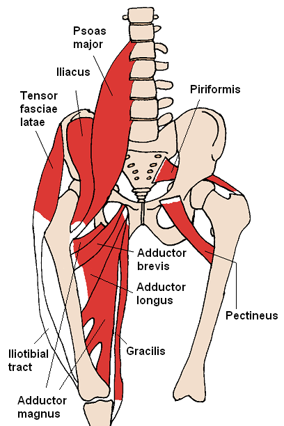
While flexion is a step forwards, extension describes the position of that hip after the other leg has taken a. Body muscle structure 12 photos of the body muscle structure body muscle chart exercises, body muscle chart for bodybuilding, body muscle names chart, body muscle ratio chart, human body muscle chart free, human muscles, body muscle chart exercises. The achilles tendon attaches the muscles of the. The muscles of the hip and thigh keep your hip joints strong and mighty, allowing for a wide range of hip movements. The hip muscle diagram below shows a number of the muscles we will be discussing in the next sections.
The muscles of the lower back, including the erector spinae and quadratus lumborum muscles, contract to extend and laterally bend the vertebral column.
Dislocation of the hip joint. • posterior • piriformis • gemellus superior • obturator internus • gemellus inferior • quadratus femoris. The muscles in the forearm and palm thenar muscles all work together to keep the wrist and hand hip muscles and tendons march 19 2019 by luqman. Hip and thigh muscles (overview diagram). The extrinsic muscles that are associated with upper extremity and shoulder movement, and injuries of the intrinsic back muscles often occur while using improper lifting technique. You can protect the back muscles by bending from the hip and. Back muscles anatomy lower back muscles anatomy human anatomy. The back's muscles start at the top of the back (named the cervical vertebrae) and go to the tailbone (also named the coccyx). Learn with flashcards, games and more — for free. The muscles of the lower back, including the erector spinae and quadratus lumborum muscles, contract to extend and laterally bend the vertebral column. Other muscles are small and cover much less space. • the sciatic nerve passes just inferior to the piriformis therefore a tight piriformis muscle my contribute to compression on the sciatic nerve. Abduction and medial rotation at the hip.
In human anatomy, the muscles of the hip joint are those muscles that cause movement in the hip. Abduction and medial rotation at the hip. Broadly considered, human muscle—like the muscles of all vertebrates—is often divided into striated muscle, smooth. The core muscles are those in the abdomen, back, and pelvis, and they also stabilize the body and assist in tasks, such as lifting weights. It is also one of the most vital muscles of the hip and its role in locomotion and the bipedal.

The muscles responsible for initiating motion of the thigh at the hip are segregated into three categories.
Sit on the floor with your legs extended straight in front of you 2. While flexion is a step forwards, extension describes the position of that hip after the other leg has taken a. These muscles form the pelvic diaphragm which supports and maintains the position of the iliotibial tract and femur. Want to learn more about it? Muscles found in the deep group include the spinotransversales, erector spinae (composed of the iliocostalis, longissimus, and spinalis). The gluteus medius, gluteus minimus, piriformis, tensor fasciae latae on the outside. Human muscle system, the muscles of the human body that work the skeletal system, that are under voluntary control, and that are concerned with movement, posture, and balance. The levator ani muscle along with a second muscle forms the pelvic floor. Learn with flashcards, games and more — for free. Common hip and back pain causes include injury to muscles from overuse disc injurydegeneration or spinal stenosis. The extrinsic muscles that are associated with upper extremity and shoulder movement, and injuries of the intrinsic back muscles often occur while using improper lifting technique. Muscles of hip bone and spine. Gluteus maximus, biceps femoris, semitendinosus, semimembranosus at the back and the.
To learn more about the lower back anatomy of the spine, please watch this video. The levator ani muscle along with a second muscle forms the pelvic floor. Dislocation of the hip joint. Lying down variation 1.lie flat on your back. Learn with flashcards, games and more — for free.

• the sciatic nerve passes just inferior to the piriformis therefore a tight piriformis muscle my contribute to compression on the sciatic nerve.
Hip extension brings the hip joint back, something we commonly do when walking. Deadlift muscles will include knee, hip, and back extensors, which primarily include the quads, glutes, and spinal erectors. The main muscles of the hip and pelvis consistsof the iliopsoas, pectinues, rectus femoris and sartorius at the front. Back muscles anatomy lower back muscles anatomy human anatomy. The gluteus medius, gluteus minimus, piriformis, tensor fasciae latae on the outside. • the sciatic nerve passes just inferior to the piriformis therefore a tight piriformis muscle my contribute to compression on the sciatic nerve. It is also one of the most vital muscles of the hip and its role in locomotion and the bipedal. It is opposite from the chest, and the vertebral column runs down. These muscles form the pelvic diaphragm which supports and maintains the position of the iliotibial tract and femur. Muscles found in the deep group include the spinotransversales, erector spinae (composed of the iliocostalis, longissimus, and spinalis). Some of these muscles are quite large and cover broad areas. Now that you watched the video, you. To learn more about the lower back anatomy of the spine, please watch this video.


Komentar
Posting Komentar